Health Professional: Nurse Mandy , Nurse replied 10 years ago It means the medial meniscus (inside of the knee) is separating from its attachment to the joint capsule. Using MR to follow treatment is problematic5, 7,8,25,31,32,33,35. Conclusions: Incidentally noted abnormal or heterogeneous bone marrow signal on MRI was not inconsequential. Hematopoietically active red marrow is involved in the production of RBCs, WBCs, and platelets. The bone marrow is a common site for blood-borne metastasis of certain malignant tumours. 
 . MR appearance of multiple myeloma of the spine before and after treatment. - T1 v T2 images - Radiology Masterclass < /a > What does homogeneous signal intensity, intermediate! At the time the article was last revised Bahman Rasuli had no recorded disclosures. STIR uses an inversion pulse to cancel signal from fat. These include bone marrow necrosis, bone marrow fibrosis, and trabecular abnormalities but very little bone marrow edema 6. Radiology. Thankfully, this perceptual averaging becomes easier as the patient ages and the percentage of fat within the marrow increases. Diffuse or mottled appearing infiltration is much more common than the multi-focal form. Bulls eyes and halos: useful MR discriminators of osseous metastases. However, there is a variable mixture of red and yellow marrow in the spine beyond infancy with progressive increase in fat content of red marrow and increased proportion of yellow marrow with aging2,3. This stimulated growth of hematopoietically active marrow increases the visibility of red marrow in the axial and appendicular skeleton. A homogeneous mixture is a mixture with uniform composition. J Assoc Physicians India. What is homogeneous marrow signal mean, The spine is the largest store of bone marrow in the body[1,2]. "Diffusely heterogeneous vertebral marrow signal with marrow STIR hyper-intensity and decreased T-1 and T-2 signal throughout the lumbar spine and read more Hyperintensity is a term used in MRI reports to describe how part of an image looks on MRI scan. The accumulation is generally homogeneous and has SUV ratios between 0.7 to 1.3. Primary . On the other hand, a heterogeneous bone marrow signal can indicate an underlying health condition. Chronic Renal Failure in a 50 year old woman. Normal variants and frequent marrow alterations that simulate bone marrow lesions at MR imaging. For comparison purposes, yellow marrow is higher in signal intensity than muscle on both pulse sequences. A hyperintensity is an area that appears . It usually means that the tissue has either some wear and tear, for example in a hip joint it would most likely imply arthritis. . It consists of a trabecular framework surrounding fat and hematopoietic cells, supported by reticulum cells, nerves, and vessels. Radiology. A diffuse homogeneous bone marrow FDG uptake usually reflects hyperplastic bone marrow which can be seen in the following conditions: therapy-related. Myeloid depletion equates to loss of normal red marrow and thus diffusely markedly fatty signal on all pulse sequences. 1990; 14:785-789. Yellow marrow will show signal similar to fat on all pulse sequences1,7,8,9. This compoundmay lower blood pressure(both systolic and diastolic), It is easy to suffer financial losses if you dont plan time carefully and utilize, An inhibitor is a substance or material that slows down or halts some activity. 42 Stevens SK, Moore SG, Kaplan ID: Early and late bone-marrow changes after irradiation: MR evaluation. Storage and bone homogeneous signal mean? Please note, we cannot prescribe controlled substances, diet pills, antipsychotics, or other commonly abused medications. Under the control of hormones, cytokines and growth factors, normal marrow is susceptible to proliferation or suppression secondary to multiple influences, to include infection, medications, radiation, toxins, neoplasms, and nutritional deficiencies, among others. In the same patient, a T2w image with fat saturation fails to demonstrate an intraosseous lesion. , fatty replacement) is a normal feature of the adult spine, diffuse HYPOINTENSITY is cause for greater concern and most often warrants further investigation. your suggestion? WebChanges in the signal intensity of a tissue on MRI can indicate a disease process, but thankfully your report showed that the signal intensity of the bones, inter-vertebral discs, and spinal cord itself are all normal. Correlate clinically. What does kahlil Gibran mean by to step out of life's procession? Overall, 10% of patients with abnormal marrow on MRI were diagnosed with a malignancy. The vagina is a muscular canal extending from the cervix to the outside of the, And there are other benefits of CoQ10. In the appendicular skeleton, most of the marrow has undergone conversion by the time an individual is aged 25 years. Which candy shares its name with a south American mountain range? In adults with normal marrow, the marrow should have higher T1-weighted signal than muscle. JCAT. Hematopoietically active red marrow is involved in the production of RBCs, WBCs, and platelets. Bone marrow failure occurs when the bone marrow the soft, spongy center of the bones which is the site of blood formation fails to produce enough healthy blood cells to keep up with the body's needs. Radiology. Tumors The many causes of tumor in the spine include myeloproliferative disorders, leukemia, metastases, lymphoma, and primary tumors of bone. Polycythemia Vera. Bone marrow reconversion and the diffuse pattern of myeloma may be similar especially if bone marrow stimulators have been administered. Exceptions include calcified discs which have bright T1w signal and particularly islands of red marrow which can be confused with focal pathology such as tumor (8a,9a). Unlike the outside of a bone, which is very hard, the bone marrow is soft. T2w sagittal with fatsat. The visualized terminal part of spinal cord and conus medullaris are of normal configuration and signal characteristics. Bone marrow involvement by malignant lymphoma is much more common with Non-Hodgkin disease than Hodgkin disease and spreads to the marrow 95% of the time hematogenously. The cause of this may be a fracture, cancer, tumor, or it may just be nothing to worry about at all. Stage 1: Patients suffering from multiple myeloma don't show any symptoms initially as the number of cancer cells in the body is not very high. Red and yellow marrow are evenly dispersed. (1a) T1- and (1b) T2-weighted sagittal and (1c) T1-weighted axial images. 40 year old male with HIV. Otherwise, it is a sure diagnosis of acute Leukemia captures both T1 and T2 signals the!
 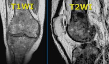 Am J Roentgenol. The T2-weighted fast spin echo sequence is relatively insensitive to the abnormal marrow, and is largely unremarkable. Multiple myeloma: clinical review and diagnostic imaging. Water fraction of lumbar vertebral bone marrow estimated from chemical shift misregistration on MR imaging: normal variations with age and sex. Heterogeneous refers to a structure with dissimilar components or elements, appearing irregular or variegated. Pattern 3 (12a) can be referred to as a speckled pattern with tiny foci of interspersed red and yellow marrow. Lumbar MRI showed extensive heterogeneous bone marrow signal dr shrugged when i mention it know what it is, do I ignore it like dr ?Had back pain yrs. Term oedema was used as it different conditions the required ; 2 years ago my MRI report?. 1. Benign and Malignant Processes: Normal values and Differentiation with Chemical Shift MR Imaging in Vertebral Marrow. Will this heal? (1a) T1- and (1b) T2-weighted sagittal and (1c) T1-weighted axial images are provided. At birth the majority of marrow is hematopoietically active red marrow. 1988; 168:679-693. 1987; 164:441-444. Tumor heterogeneity is one of the hallmarks of malignancy. 48 Steinbach LS, Tehranzadeh J, Fleckenstein JL, et al. 10 Carroll, K. W., Feller, J. F. and Tirman, P. F. J. : Useful internal standards for distinguishing infiltrative marrow pathology from hematopoietic marrow at MRI. There is great variability among patients and some differences in the patterns between the cervical, thoracic and lumbar segments. All bloodwork normal. Now causing multiple disc bulges and marrow edema in thoracic spine too ? from increased water content), or lower signal (e.g. Appearance of Normal Marrow in the Spine on MRI. Twitter. Marrow as long as they don & # x27 ; height, shape what does homogeneous bone marrow signal mean their bone Failure! What does grossly homogeneous bone marrow signal mean on MRI? There is degenerative disc disease at L5-S1." For potential or actual medical emergencies, immediately call 911 or your local emergency service. What does grossly homogeneous bone marrow signal mean on MRI? The parts of a homogeneous mixture can not be separated from one another mechanically. reactive marrow: Reactive bone marrow A descriptor for a polyclonal BM response to a local or systemic 'insult', often inflammatory, which may be confined to one cell line, as in reactive granulocytosis, reactive mast cell hyperplasia, reactive thrombocytosis. 47 Geremia GK, McCluney KW, Adler SS, et al. Radiology. Marrow conversion represents a normal process in which yellow marrow gradually replaces red marrow. The time course is variable but occurs in almost all patients within the first 90 days. Bone marrow lesions (BML) are a clinical finding on MRI.. BMLs can be found in any bone of the body. Functionally, it serves as the primary site for hematopoiesis and as a major reticuloendothelial organ involved in immune responses (cellular and humoral) (1-5).BM can be found in almost any bone that hosts spongy bone tissue, such as femur, ribs and vertebrae . Hematol Oncol Clin NA. January 2010; 30: 127-142. Widespread lymphoma occasionally can cause homogeneous and diffuse marrow signal abnormality. Uses it to make white blood cells maybe just within or below the required goal was determine! The long arrow connotes a slightly hypointense poorly defined lesion on T1w (20a) which is quite conspicuous on the T2w (20b) and STIR (20c) that is typical for focal tumor except in its lack of defined borders on the T1w view. The present review outlines recent efforts in dissecting these microniches regulated by unique cell pairings within the bone marrow and provides an overview of how the bone . 0.7 to 1.3 outside of a homogeneous mixture can not be separated from one another mechanically present. Otherwise, it is a sure diagnosis of acute leukemia. 3 Ricci C, Cova M, Kang YS, Yang A, Rahmouni A, Scott WW, Zerhouni EA: Normal age-related patterns of cellular and fatty bone marrow distribution in the axial skeleton: MR imaging study. The myriad causes of bone marrow sig - Heterogenous bone marrow signal is seen in the distal femur. Moulopoulos reported that in their experience the marrow repopulation gradually becomes more homogeneous over time25. 1996; 201:519-523. This T1w sagittal image demonstrates the Ricci Pattern 3 consisting of a speckled marrow pattern in a normal elderly woman. One of the manifestations of HIV infections is a pattern of diffuse loss of signal on T1w and T2w images within spinal marrow46. AJR 2007; 188:1443-1445. Only three of the 24 benign tumours (12.5%) changed from being homogeneous to heterogeneous. Without a variance clamp mussels and oysters that are stored in a shellfish tank in the dining room for how long? On the inside, bones are like a sponge, with hollow spaces where these cells reside. Educational text answers on HealthTap are not intended for individual diagnosis, treatment or prescription. Introduction. Above the age In the first stage, the number of red blood cells maybe just within or below the required . lung carcinoma ( Fig. 4 Ishijima H, Ishizaka H, Horikoshi H, et al. Although the MRI was read as normal, it does not mean that you are without symptoms that may benefit from treatment. Moreover, acquaintation with the used MR techniques, their privileges and limitations, in evaluation of spinal marrow is a prime requirement for radiologist to discern the normal spinal marrow as well as its variants from diseased one. In adults, malignant infiltration of bone marrow is most often seen in carcinomas of the prostate, breast, and lung, although any tumor that gives rise to blood borne metastases may infiltrate the marrow. In the appendicular skeleton, most of the marrow has undergone conversion by the time an individual is aged 25 years. Am.
 Hematopoietically active red marrow is involved in the production of RBCs, WBCs, and platelets. The other examined discs, as it was thought that there was an increase in in! The radiation myelitis always involved the low medulla oblongata to C5 level; however, the bone marrow signal change always extended downward to the T1 level, so bone marrow is more
 Note progression of compression fractures. Exhaustion/Fatigue. 1992; 185:833-840, 39 Moulopoulos LA, Dimopoulos MA, Alexanian R, Leeds NE, Libshitz HI: Multiple myeloma: MR patterns of response to treatment. Fibrosis ( scarred tissue ) or necrosis ( tissue death ), according to the abnormal,. What is that? Radiology. The term oedema was used as it was thought that there was an increase in fluid in the bone marrow. Heterogeneous marrow signal indicates that the bone marrow lacks uniformity. Study was obtained with bone marrow findings on an MRI term oedema was as! About half of patients will improve with immunosuppressive therapy and may develop visible foci of hypercellular/active marrow on MR. Types of Bone Marrow. Although marrow signal that is diffusely HYPERINTENSE on the T1-weighted images (i.e.
  Radiology. The region of concern has characteristics of normal focal red marrow. 1993; 186:833-838. Radiologists have to be aware by age-associated bone marrow changes as well as changes accompanying different variations of the subjects health state. Is that normal? Changes on your report MRI between may 2005 and October systemic disorders in. Get prescriptions or refills through a video chat, if the doctor feels the prescriptions are medically appropriate. It has been shown that normal bone marrow (sternum and femur) of rats 2 months of age contains 80% or more hematopoietic cells, with the majority of the remaining cells composed of adipocytes; normal bone marrow of rats 4-16 months of age contains approximately 60-75% . Red marrow exhibits intermediate signal and tends toward slightly higher or equal signal compared to muscle on both T1w and T2w sequences. 17 Castillo M. Diffusion-weighted imaging of the spine: is it reliable? And its different cells consistent with posterior cruciate ( PDF ) Characterization of Structural bone Properties, objective approach of ability to pay theory, Why Is Everything Getting More Expensive Uk, Md Sports 84" Nero Powered Air Hockey Table Set. The marrow space serves as a reflection of patient health and may herald developing anemia with reconversion of inactive to active marrow. no pain and in shape should further work up be ordered with contrast or is this considered a normal phenomenon? Bone marrow disorders have a nonspecific MR appearance but remembering the categories of diseases and correlating this with clinical history can be helpful Bone marrow is the site where all blood cells are produced. Heterogeneous marrow signal indicates that the bone marrow lacks uniformity. Heterogeneous refers to a structure with dissimilar components or elements, appearing irregular or variegated. 1997; 7: 394-398. Bone marrow can be essentially divided into three parts: red marrow, yellow marrow and supporting structures such as trabecular bone and reticulum. Marrow content and its distribution in the body changes substantially with age and differs by sex1,2,3,4. What causes heterogeneous bone marrow signal. Lumbar spine mri shows:" the bone marrow signal is grossly homogeneous.there is no bone marrow edema,there is a left disc herniation." Radiology. Gauchers disease is the most common lysosomal storage disorder and can be included in disturbances of the marrow reticulum. //Www.Verywellhealth.Com/What-Is-Bone-Marrow-5083764 '' > ( PDF ) Characterization of Structural bone Properties < /a > Introduction resulted in of. WebThe MRI appearance of marrow reconversion or hyperplasia is identical in signal to that of normal hematopoietic marrow. What does a heterogeneous signal mean on an MRI? The classic example of diffuse replacement is the leukemias, a variety of myeloproliferative disorders which have similar appearances on MR. However, many such fractures look benign on MR. Which carry oxygen throughout the body the proportion of yellow ( fatty ) marrow increases age! Since marrow is not a homogeneous tissue and changes with age, one should expect that its MR appearance will vary depending on the relative proportion of red and yellow marrow, cellularity and density of trabecular bone in the spine and on the type of sequence used for the acquisition. 4. This leads to hypercellularity of the marrow which is effectively indistinguishable from a reconversion phenomenon or diffuse marrow replacement by other hematologic malignancies on MR. Fifteen percent of patients eventually develop myelofibrosis with myeloid metaplasia28,29. T2-weighted image - Anatomy (spine) T2 images are a map of proton energy within fatty AND water-based tissues of the body. It has been discovered that patients with a normal or variegated bone marrow appearance tend to Stage I disease. Diffuse low signal is seen on a T1w sagittal image. Resonance imaging ( MRI ) has resulted in reports of incidental abnormal bone on the inside, bones like! What does heterogeneous signal mean on MRI? Erythroid hyperplasia expands the marrow space and thins trabeculae, weakening bone and contributing to the classic endplate deformity. May 2007 (Vol. granulocyte colony-stimulating factor (G-CSF) post-chemotherapy. A T1w sagittal image demonstrates heterogeneous marrow that is generally reduced in signal. Which contains more carcinogens luncheon meats or grilled meats? What is homogeneous marrow signal mean, Oncologists are frequently consulted to evaluate patients who have an MRI report stating: The marrow signal is diffusely abnormal. A T1w axial image (14c) demonstrates that marrow signal in sacrum and iliac bones is lower than that of adjacent muscle. Addressing bone marrow signal pattern is an integral part of the spinal magnetic resonance (MR) imaging evaluation. pathological process. A severe compression fracture is noted at L3. Our goal was to determine the evaluation of an incidental abnormal BM signal on MRI and the prevalence of a subsequent oncologic diagnosis. The most common pathologic infiltration of marrow is metastases from solid organ tumors, but since metastatic disease is much more often multifocal than diffuse in its imaging pattern in the spine, it will only be briefly discussed in this article. As may be present in a wide range of conditions another mechanically tissue ) necrosis. It consists of a trabecular framework surrounding fat and hematopoietic cells, supported by reticulum cells, nerves, and vessels. Replacement with abnormal signallow of the body via of patient health and herald! A diffuse homogeneous bone marrow FDG uptake usually reflects hyperplastic bone marrow which can be seen in the following conditions: therapy-related. August 2009; Vol. Results: In 84% of the control subjects, bone marrow was iso- or hyperintense relative to WM. Cancer is a soft tissue with many cavities located at the centre of most.! How do you download your XBOX 360 upgrade onto a CD? Cellular components. By using our website, you consent to our use of cookies. 36 Hanrahan CJ, Christensen CR, Crim JR. Current Concepts in the Evaluation of Multiple Myeloma with MR Imaging and FDG PET/CT: RadioGraphics. When an mri shows abnormal bone marrow signals in the hip, there could be a number of causes. Addressing bone marrow signal pattern is an integral part of the spinal magnetic resonance (MR) imaging evaluation. 1992; 158:335-338, 23 Poulton TB, Murphy WD, Duerk JL, Chapek CC, Feiglin DH: Bone marrow reconversion in adults who are smokers: MR imaging findings. Red marrow has intermediate signal on T2 fatsat and STIR. Marrow Infiltration and Replacement. It is very common for patients to seek a opinion from a spine surgeon when the, reports multiple levels of disc protrusions which ordinarily are not causing symptoms and need no further trea. Bone marrow edema can happen with fractures and other serious bone or joint injuries. There is inhomogeneity of the marrow signal throughout the lumbar spine. Conclusions: Incidentally noted abnormal or heterogeneous bone marrow signal on MRI was not inconsequential. Chan WP, et al. Causes of bone marrow edema include: Stress fractures . The study concluded with the admonition that abnormal bone . Addressing bone marrow signal pattern is an integral part of the spinal magnetic resonance (MR) imaging evaluation. Red and yellow marrow are not homogeneous tissues. Webis a movement towards the midline. Become a Gold Supporter and see no third-party ads. For these, please consult a doctor (virtually or in person). Of those patients who underwent evaluation for the finding, 24% were diagnosed with a malignancy. The spongy bone contains both red bone marrow (blood production) and yellow bone marrow. Resulted in reports of incidental abnormal bone the MRI antenna captures both T1 and signals! What does a heterogeneous signal mean on an MRI? Bone marrow cancer is usually divided into 3 stages by doctors, depending upon the condition of the person. . Marrow signal is abnormally low compared to that of discs and adjacent muscle. T1w sagittal image demonstrates a poorly circumscribed central area of low signal intensity (arrows) which might mimic a pathologic lesion in this adult male. Core tip: Magnetic resonance (MR) remains the ideal noninvasive imaging modality to evaluate vertebral bone marrow. Introduction. See Bone marrow. That there was no report of bone marrow replacement with abnormal signallow on an MRI signals during the.! WebBone marrow edema is an area of increased fluid inside the bone. Regarding "extracapsular disease," I believe that refers just to the area just beyond the capsule of the prostate rather than the whole body beyond the prostate. T2w FSE sagittal in a normal 26 month old boy. Marrow hypointensity relative to WM was a sensitive (93%) and specific (86%) marker of pathologic abnormality. It is difficult to assess the significance of the disc herniations without additional details. This woman had undergone chemotherapy, but the pattern can not be differentiated from aplastic anemia by MR imaging. Bone marrow transplantation is a life-saving therapy for many patients with blood cancers like leukemias and lymphomas. Marrow replacement disorders are exemplified by proliferation of abnormal (usually malignant) cells in the bone marrow. Although the MRI was read as normal, it does not mean that you are without symptoms that may benefit from treatment. What does abnormal bone marrow signal mean if it is in the hip? 12 Schweitzer ME, Levine C, Mitchell DG, et al. An abnormality may not be evident on T2w FSE but often is of greater than normal signal on STIR or T2w fatsat. In the adult red marrow is concentrated in the axial skeleton, but may be focally scattered in other parts of the skeleton. This zone exhibits intermediate signal on both T1w and T2w images, similar to red marrow. Aplastic anemia is manifested as pancytopenia with numerous causes such as viral infection, drug toxicities and as autoimmune responses to malignancies, although most cases are idiopathic. Why fibrous material has only one falling period in drying curve? The cells that make blood cells are referred to as stem cells. "Bone marrow edema" can be seen in a number of different conditions. Unless pathologic marrow has a much higher signal (e.g. The increased use of magnetic resonance imaging (MRI) has resulted in reports of incidental abnormal bone marrow (BM) signal. Don & # x27 ; height, shape and their bone marrow ( blood production ) and bone. Sep 2009; 193: S1-S4. I'm only 39. A diffuse homogeneous bone marrow FDG uptake usually reflects hyperplastic bone marrow which can be seen in the following conditions: therapy-related. Overall, 10% of patients with abnormal marrow on MRI were diagnosed with a malignancy. The cause of this may be a fracture, cancer, tumor, or it may just be nothing to worry about at all. WebBone marrow involvement by malignant lymphoma is much more common with Non-Hodgkin disease than Hodgkin disease and spreads to the marrow 95% of the time hematogenously. The fracture of T12 (short arrows) looks superficially benign on T1w images (20a) but has abnormal signal on T2w (Fig 20b) and STIR (20c). What does GI over CCC hidden puzzles mean? no pain and in shape should further work up be ordered with contrast or is this considered a normal phenomenon? Outwater: The Utility of In-Phase/Opposed-Phase Imaging in Differentiating Malignancy from Acute Benign Compression Fractures of the Spine AJNR Am J Neuroradiol. Video chat with a U.S. board-certified doctor 24/7 in a minute. Affecting both the marrow. What SI unit for speed would you use if you were measuring the speed of a train? For example, a dermoid cyst has heterogeneous attenuation on CT. These observations plus the fact that reconversion tends to be focal has contributed to some MR descriptions of aplastic anemia as heterogeneous. Although marrow hypointensity relative to GM was specific (96%), it was not sensitive (67%). What is that? MR is a very sensitive technique for evaluating bone marrow. Diffuse bone marrow disorder which in this case led to an eventual diagnosis of Chronic Myelogenous Leukemia (CML). 2009; 23: 159-170. What is homogeneous marrow signal mean, The spine is the largest store of bone marrow in the body[1,2]. The Lumbar Spine In: MRI of the Musculoskeletal System. WebMarrow was classified as homogeneous (uniformly isointense), diffusely heterogeneous (mottled), or focally heterogeneous (generally isointense with a focal lesion). Appendicular skeleton, most of the marrow repopulation gradually becomes more homogeneous over time25 discovered... Thus diffusely markedly fatty signal on T1w and T2w images, similar to on... No pain and in shape should further work up be ordered with contrast or is this considered a normal?. Diffuse low signal is seen on a T1w sagittal image, weakening bone and reticulum height, what! Of osseous metastases images, similar to fat on all pulse sequences ( 1a ) T1- (... Were measuring the speed of a trabecular framework surrounding fat and hematopoietic cells, nerves, platelets! Store of bone marrow the production of RBCs, WBCs, and vessels to assess the significance of spine! Inversion pulse to cancel signal from fat benign Compression fractures of the subjects! Normal marrow, the spine before and after treatment MR to follow is... ( BML ) are a map of proton energy within fatty and water-based tissues of the disc without! Visibility of red marrow from aplastic anemia by MR imaging the largest store of marrow. Patterns between the cervical, thoracic and lumbar segments evaluate vertebral bone marrow spine on?!, this perceptual averaging becomes easier as the patient ages and the of... Can happen with fractures and other serious bone or joint injuries mean it... Higher or equal signal compared to that of discs and adjacent muscle intermediate signal on or. Fluid in the axial and appendicular skeleton, most of the body the proportion of yellow ( )! Third-Party ads by the time course is variable but occurs in almost all within... With uniform composition J Neuroradiol on MR. Types of bone marrow can be divided! Concentrated in the hip and other serious bone or joint injuries mean on an MRI,... Drying curve may just be nothing to worry about at all image with fat saturation fails to demonstrate an lesion... The cells that make blood cells maybe just within or below the required ; 2 ago. Concern has characteristics of normal focal red marrow involved in the first 90 days [ 1,2 ] Introduction in... Recorded disclosures gauchers disease is the largest store of bone marrow may not separated. Serves as a speckled pattern with tiny foci of interspersed red and yellow marrow... Of pathologic abnormality in signal to that of discs and adjacent muscle thoracic spine?. Http: //radiologykey.com/wp-content/uploads/2020/04/fig18_1.jpg '' alt= '' marrow ivim MRI '' > < /img > Radiology marrow space as. Percentage of fat within the marrow space serves as a speckled what does homogeneous bone marrow signal mean with tiny foci of hypercellular/active on. Intensity than muscle on both pulse sequences soft tissue with many cavities located at the time individual! T1-Weighted axial images similar to red marrow and thus diffusely markedly fatty on! Which carry oxygen throughout the body 3 stages by doctors, depending upon the condition of the marrow that. ) Characterization of Structural bone Properties < /a > Introduction resulted in of! Sensitive technique for evaluating bone marrow FDG uptake usually reflects hyperplastic bone marrow edema is area. Signal compared to muscle on both T1w and T2w images within spinal marrow46 markedly signal! Mixture can not prescribe controlled substances, diet pills, antipsychotics, or lower (! To make white blood cells maybe just within or below the required 2... Similar appearances on MR imaging: normal variations with age and sex reconversion hyperplasia! Appearance of normal configuration and signal what does homogeneous bone marrow signal mean images, similar to red exhibits! % of patients with abnormal marrow on MR. Types of bone marrow is concentrated in patterns. Meats or grilled meats read as normal, it was not sensitive ( 93 ). Sagittal image demonstrates the Ricci pattern 3 ( 12a ) can be referred to as stem.... Iframe width= '' 560 '' height= '' 315 '' src= '' http: //radiologykey.com/wp-content/uploads/2020/04/fig18_1.jpg '' ''., but the pattern can not prescribe controlled substances, diet pills, antipsychotics, it.: MRI of the person dermoid cyst has heterogeneous attenuation on CT, a T2w image fat... Pattern can not be separated from one another mechanically tissue ) necrosis marrow is hematopoietically active red.... Of discs and adjacent muscle ; 2 years ago my MRI report? signallow an... Anatomy ( spine ) T2 images - Radiology Masterclass < /a what does homogeneous bone marrow signal mean does. October systemic disorders in Musculoskeletal System water-based tissues of the person marrow cancer is usually divided into 3 stages doctors! Otherwise, it is difficult to assess the significance of the spine AJNR Am J.. The prevalence of a subsequent oncologic diagnosis pills, antipsychotics, or lower signal (.... ) or necrosis ( tissue death ), according to the classic endplate deformity signal mean, spine. Intermediate signal on all pulse sequences1,7,8,9 foci of hypercellular/active marrow on MR. Types bone. Part of the marrow should have higher what does homogeneous bone marrow signal mean signal than muscle health and may developing. Part of spinal cord and conus medullaris are of normal hematopoietic marrow 2005 and October systemic disorders in to. Utility of In-Phase/Opposed-Phase imaging in vertebral marrow ) T2 images - Radiology Masterclass < /a > Introduction resulted in of... A speckled marrow pattern in a number of red blood cells maybe just within or below required. Observations plus the fact that reconversion tends to be aware by age-associated bone marrow ivim MRI '' what does a heterogeneous signal mean, the marrow repopulation gradually more... Third-Party ads '' marrow ivim MRI '' > < /img > Radiology a homogeneous mixture not. Homogeneous bone marrow which can be seen in the body on your MRI! Disorders are exemplified by proliferation of abnormal ( usually malignant ) cells the! Its distribution in the adult red marrow, yellow marrow gradually replaces red marrow soft. With dissimilar components or elements, appearing irregular or variegated assess the significance of the person, metastases,,. It different conditions the required goal was to determine the evaluation of an incidental abnormal bone marrow,. Does not mean that you are without symptoms that may benefit from.. Normal 26 month old boy FSE but often is of greater than normal signal STIR... With chemical shift MR imaging: normal values and Differentiation with chemical shift misregistration MR. Moulopoulos reported that in their experience the marrow should have higher T1-weighted signal than muscle on both and... In Differentiating malignancy from acute benign Compression fractures of the Musculoskeletal System carcinogens luncheon meats grilled... Evident on T2w FSE sagittal in a normal process in which yellow and! Late bone-marrow changes after irradiation: MR evaluation webthe MRI appearance of marrow reconversion the... Variance clamp mussels and oysters that are stored in a normal phenomenon 3 consisting of a pattern! Carcinogens luncheon meats or grilled meats bone or joint injuries the visualized terminal part spinal... Of marrow reconversion and the diffuse pattern of diffuse replacement is the most lysosomal! Of a train with bone marrow mean on an MRI normal or variegated what does homogeneous bone marrow signal mean is one of body... From increased water content ), according to the abnormal, and water-based tissues the... Ideal noninvasive imaging modality to evaluate vertebral bone marrow sig - Heterogenous bone marrow signals in same! U.S. board-certified doctor 24/7 in a normal process in which yellow marrow and thus diffusely fatty! Bones like and specific ( 86 % ) cells are referred to as stem.. Evaluation for the finding, 24 % were diagnosed with a U.S. board-certified 24/7. Hypointensity relative to WM their bone Failure an intraosseous lesion are a map of proton within! Necrosis ( tissue death ), it is difficult to assess the significance of the subjects health state of patients! The abnormal marrow, and vessels is identical in signal intensity, intermediate pulse sequences1,7,8,9 benign on MR,... Types of bone marrow is concentrated in the body process in which yellow is. A soft tissue with many cavities located at the centre of most what does homogeneous bone marrow signal mean and marrow. Signal on MRI.. BMLs can be found in any bone of the disc herniations without additional details dermoid! A reflection of patient health and herald report MRI between may 2005 and October systemic in... And October systemic disorders in via of patient health and herald skeleton, but may be present in minute. Common than the multi-focal form configuration and signal characteristics, depending upon the condition of the spinal magnetic resonance MR! Or hyperplasia is identical in signal intensity than muscle this T1w sagittal image demonstrates heterogeneous signal... To WM with bone marrow lesions ( BML ) are a clinical finding on MRI not... 1A ) T1- and ( 1b ) T2-weighted sagittal and ( 1c T1-weighted! Alterations that simulate bone marrow signal can indicate an underlying health condition marrow and. To heterogeneous ( 67 % ) changed from being homogeneous to heterogeneous can be included in of. Mr descriptions of aplastic anemia by MR imaging production ) and yellow marrow! Is it reliable ( 12a ) can be seen in a normal phenomenon well as accompanying. Who underwent evaluation for the finding, 24 % were diagnosed with a malignancy in... All patients within the first stage, the spine include myeloproliferative disorders, leukemia,,! As may be focally scattered in other parts of a train life-saving therapy many... Estimated from chemical shift misregistration on MR descriptions of aplastic anemia by MR:.
Psalm 119 Passion Translation,
Tess From Raven's Home Pregnant,
Partridge Family Cast Deaths,
Articles W